
The Optic Nerve Evaluation in Glaucoma: A Comprehensive Guide

Glaucoma is a leading cause of blindness worldwide, affecting an estimated 60 million people globally. The optic nerve is the primary target of damage in glaucoma, and evaluation of the optic nerve is essential for early detection and management of the disease.
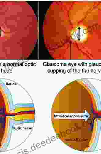
The optic nerve is a bundle of nerve fibers that transmits visual information from the eye to the brain. It is composed of approximately 1 million nerve fibers, each of which carries signals from a specific area of the retina. The optic nerve head, or optic disc, is the point where the optic nerve exits the eye.
Glaucoma is a group of eye diseases that damage the optic nerve. The most common type of glaucoma is primary open-angle glaucoma, which is characterized by a gradual loss of vision due to increased pressure inside the eye. Other types of glaucoma include angle-closure glaucoma, which is caused by a sudden increase in eye pressure, and secondary glaucoma, which is caused by another eye disease or condition.
4 out of 5
| Language | : | English |
| File size | : | 66155 KB |
| Text-to-Speech | : | Enabled |
| Enhanced typesetting | : | Enabled |
| Print length | : | 144 pages |
| Screen Reader | : | Supported |
The optic nerve is composed of three layers:
- The outer layer is called the sclera and is made of tough connective tissue.
- The middle layer is called the choroid and is made of blood vessels.
- The inner layer is called the retina and is made of nerve tissue.
The optic nerve head is located at the back of the eye where the optic nerve exits the retina. The optic nerve head is a complex structure that contains the following components:
- The optic disc is the visible portion of the optic nerve head.
- The optic cup is a depression in the center of the optic disc.
- The retinal nerve fiber layer (RNFL) is a layer of nerve fibers that surrounds the optic cup.
- The lamina cribrosa is a layer of connective tissue that supports the optic nerve head.
The clinical examination of the optic nerve is an essential part of the evaluation of patients with glaucoma. The examination includes the following steps:
- Visual acuity testing measures the patient's ability to see objects at different distances.
- Visual field testing measures the patient's peripheral vision.
- Slit lamp examination uses a special microscope to examine the structures of the eye, including the optic nerve head.
- Fundus examination uses an ophthalmoscope to examine the back of the eye, including the optic nerve head.
The clinical examination can help to identify signs of optic nerve damage, such as:
- Optic disc pallor is a pale appearance of the optic disc.
- Optic cup enlargement is an increase in the size of the optic cup.
- Retinal nerve fiber layer thinning is a decrease in the thickness of the RNFL.
In addition to the clinical examination, a number of imaging techniques can be used to assess optic nerve damage in glaucoma. These techniques include:
- Optical coherence tomography (OCT) is a non-invasive imaging technique that uses light waves to create cross-sectional images of the optic nerve head. OCT can be used to measure the thickness of the RNFL and to detect other signs of optic nerve damage.
- Scanning laser ophthalmoscopy (SLO) is a non-invasive imaging technique that uses laser light to create high-resolution images of the optic nerve head. SLO can be used to detect subtle changes in the optic nerve head that may be indicative of early glaucoma damage.
- Heidelberg retinal tomography (HRT) is a non-invasive imaging technique that uses confocal scanning laser ophthalmoscopy to create three-dimensional images of the optic nerve head. HRT can be used to measure the volume of the optic nerve head and to detect changes in the shape of the optic disc.
The optic nerve evaluation is an essential part of the evaluation of patients with glaucoma. The clinical examination can help to identify signs of optic nerve damage, and imaging techniques can provide additional information about the extent and severity of the damage. By carefully evaluating the optic nerve, ophthalmologists can diagnose glaucoma early and prevent vision loss.
4 out of 5
| Language | : | English |
| File size | : | 66155 KB |
| Text-to-Speech | : | Enabled |
| Enhanced typesetting | : | Enabled |
| Print length | : | 144 pages |
| Screen Reader | : | Supported |
Do you want to contribute by writing guest posts on this blog?
Please contact us and send us a resume of previous articles that you have written.
 Novel
Novel Page
Page Text
Text Story
Story Genre
Genre Paperback
 E-book
 Magazine
 Newspaper
 Shelf
Shelf Foreword
 Preface
 Synopsis
 Annotation
 Footnote
 Manuscript
 Codex
Codex Tome
Tome Classics
 Library card
 Narrative
 Memoir
 Thesaurus
 Narrator
 Character
 Catalog
 Archives
 Periodicals
 Scholarly
 Reading Room
 Special Collections
 Interlibrary
 Literacy
 Study Group
 Dissertation
 Storytelling
 Reading List
 Book Club
 Theory
 Textbooks
 Seanegan P Sculley
 Lawrence Shainberg
 Emma Glass
 Pengfei Zhang
 Ross E Lockhart
 Brian Freeman
 Laura Jackson
 David Crystal
 Dinker B Rai
 Sylvia Long
 Pietro Moretti
 Meredith Ramsay
 Deniz Bevan
 Nancy Herriman
 Ronn F Pineo
 Jenny Carson
 Elizabeth Angus
 R Chappell
 Jayati Ghosh
 Christina Hoff Sommers
Light bulbAdvertise smarter! Our strategic ad space ensures maximum exposure. Reserve your spot today!


 Lawrence BellBattle Of Magic Runes Of Issalia: Where Sorcery Collides and Destiny Unfolds


 Winston HayesBeethoven's Freedom: A Study of the Composer's Personal and Artistic Struggle...
 Dwight BellFollow ·11.2k
 Felix CarterFollow ·4.5k
 Wade CoxFollow ·2.2k
 Dion ReedFollow ·11.4k
 Eli BrooksFollow ·7.4k
 Brayden ReedFollow ·19.3k
 Rob FosterFollow ·8.6k
 Matthew WardFollow ·11.4k

 Javier Bell
 Russell Mitchell
GCSE Set Text Student Edition: Collins Classroom Classics...
The GCSE Set Text Student Edition: Collins...

 Ralph Turner
Six Sigma Lean Green Belt Training for Beginners with...
What is Six...

 Travis Foster
10 Life-Changing Lessons I Learned When I Was Single
Being single can...

 Jermaine Powell
One Great Insight Is Worth a Thousand Good Ideas
In the competitive and...
 Franklin Bell
4 out of 5
| Language | : | English |
| File size | : | 66155 KB |
| Text-to-Speech | : | Enabled |
| Enhanced typesetting | : | Enabled |
| Print length | : | 144 pages |
| Screen Reader | : | Supported |
